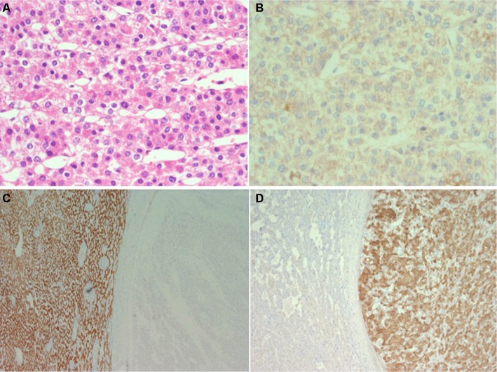
Figure 1.
Eighty-year-old woman with a large intrahepatic tumor in the right lobe suspect for hepatocellular carcinoma.
Notes: Histological examination of the resected specimen shows (A; H&E) that the tumor is composed of solid sheets or trabeculae of medium to large size cohesive cells with abundant eosinophilic cytoplasm resembling hepatocytes (hepatoid). Immunohistochemistry shows that the tumor cells do not form canaliculi (B; CEA, polyclonal antibody) and do not stain for Hep-Par 1 (C; background liver on the left and tumor on the right side of the figure) but stain for vimentin (D; same field as C) melan-A, inhibin, synaptophysin and calretinin. The final diagnosis was of liver infiltration by adrenal cortical carcinoma.

