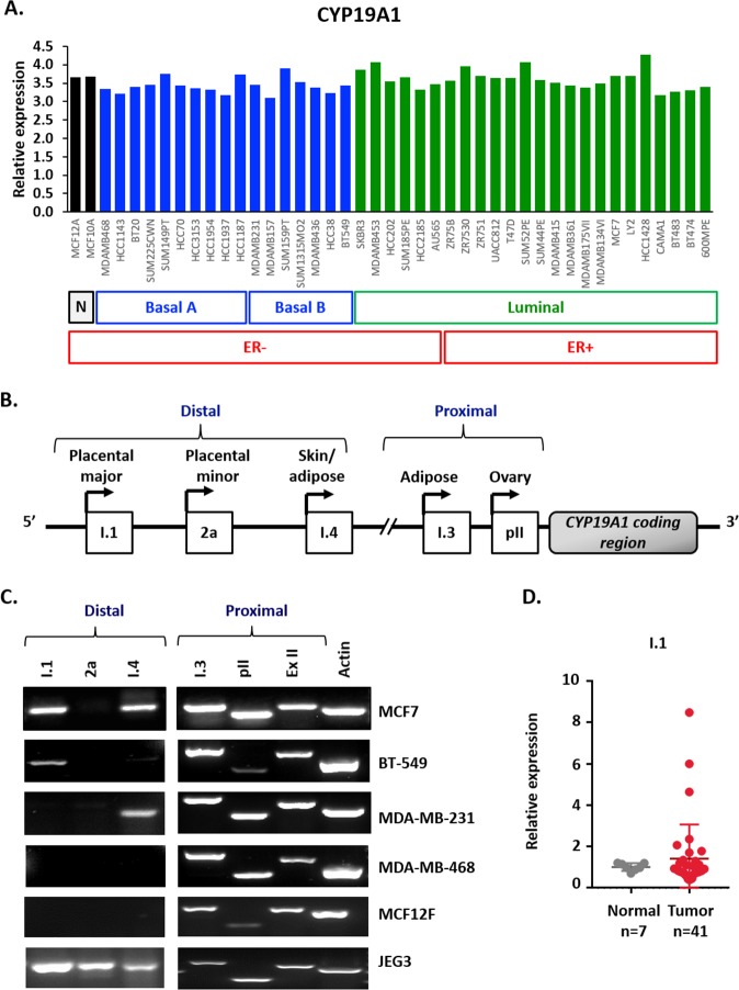
Figure 1. Multiple aromatase transcripts are expressed in multiple cancer cell lines, and the placental aromatase transcript is expressed in breast cancer tissues.
(A) Relative aromatase RNA expression in breast cancer cell lines using Heiser RNASeq data downloaded from UCSC Xena platform. (B) Schematic representation of the different tissue-specific CYP19A1 promoters: I.1, 2a, I.4, I.3 and pII. (C) Identification of aromatase active promoters in breast cancer cell lines (MCF7, MDA-MB-231, BT-549 and MDA-MB-468), non-cancer breast cell line (MCF12F) and placental cell line (JEG3) by end-point PCR. Amplification of three distal promoters, between 93 to 73Kb from ATG: placental aromatase transcripts (I.1, 2a) and skin/ adipose tissue transcript (I.4), two proximal, 0.2 Kb from ATG or less: adipose/breast cancer (I.3) and ovary/breast cancer (pII), exon II (ExII) common in all aromatase transcripts and beta-actin as control. (D) Expression of I.1 promoter in breast cancer TissueScan array (Origene) by real time qPCR, represented as average fold change of I.1 promoter respect to beta-actin and normalized to normal tissue.

