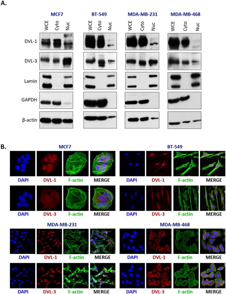
Figure 2. DVL proteins are localized in the nucleus and cytoplasm of different breast cancer cells.
(A) Nuclear and cytoplasmic extracts from four breast cancer cells (MCF7, BT-549, MDA-MB-231, and MDA-MB-468) were analyzed using Western blots. The blots were probed with DVL-1 and DVL-3 antibodies. Lamin was used as a control for nuclear extract and GAPDH was used as a control for cytosolic proteins. (B) Immunofluorescence was performed to analyze DVL proteins localization in MCF7, BT-549, MDA-MB-231, and MDA-MB-468 cells. The cells were probed with DVL-1 and DVL-3 antibodies (red). The nucleus was stained with DAPI (blue) and the actin filaments (green) were stained with Phalloidin.

