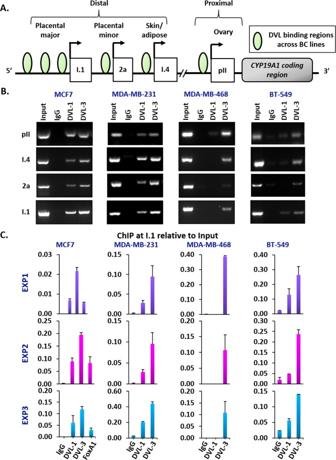
Figure 3. DVL family members bind to multiple CYP19A1 promoters.
(A) Schematic representation of different tissue-specific CYP19A1 promoters located proximally (pII, and I.3) or distally (I.4, 2a, and I.1) with respect to the coding region. The green circles represent the genomic region bound by DVL proteins in different breast cancer cells (B) Three independent ChIP experiments for IgG, DVL-1 and DVL-3 were performed in MCF7, MDA-MB-231, MDA-MB-468 and BT-549 cells. Occupancy of DVL at four tissue-specific promoters of CYP19A1 gene (pII, I.4, 2a, & I.1) were analyzed by end-point PCR. (C) Three independent ChIP-qPCR experiments at I.1 promoter for IgG, DVL-1, DVL-3 and FOXA1 were performed in MCF7 and for IgG, DVL-1 and DVL-3 in MDA-MB-231, MDA-MB-468 and BT-549 cells. error bars = std dev of triplicates.

