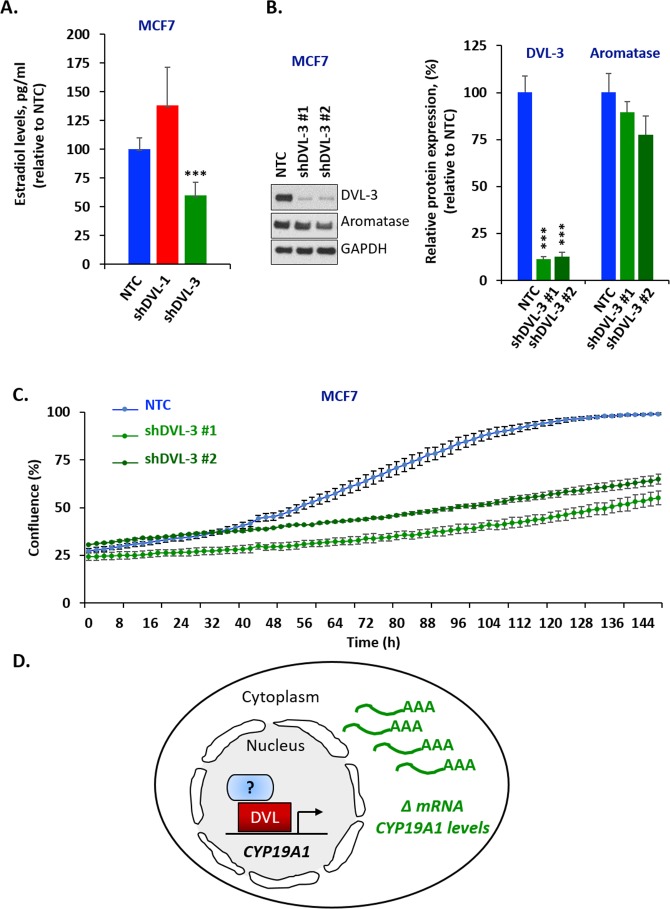
Figure 5. DVL loss of function alters estrogen levels and cell proliferation.
(A) Estradiol levels of MCF7 cells expressing stable knockdown of DVL-1 (shDVL-1) and DVL-3 (shDVL-3) and non-target control (NTC) treated with 10nM androstenedione for two days. Data are representative of 5 independent experiments carried out in triplicate with std dev, **, p= 0.0008. (B) Whole cell extracts from MCF7 NTC, MCF7 shDVL-3 #1 and MCF7 shDVL-3 #2 where analyzed using Western blots. The blots were probed with DVL-3, aromatase and GAPDH antibodies. (C) Time course of growth curve of MCF7 cells expressing stable knockdown of DVL-3 (shDVL-3 #1 and shDVL3 #2) and non-target control (NTC) cell proliferation was measured as percent confluence from phase-contrast images. Plot shows mean and SEM. Data are representative of 3 independent experiments carried out in octuplicate, *** p<0.001 after 70 h. (D) Schematic representation of DVL proteins binding to CYP19A1 promoter region and regulating its mRNA level.

