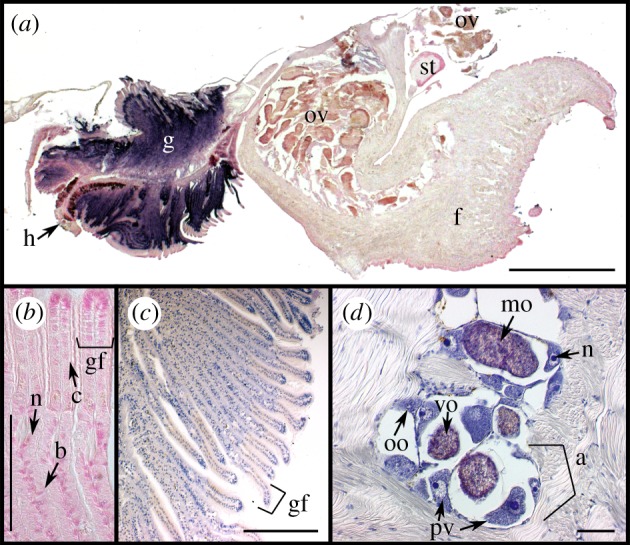
Figure 1.

Tissue ultrastructure of female S. velum imaged with differential interference contrast (DIC) microscopy. (a) Section of whole S. velum removed from shell hybridized with the symbiont probe Svsym47 (purple) and counterstained in nuclear fast red (pink/red). (b) Close-up of gill filaments stained with nuclear fast read. (c,d) Haematoxylin eosin (HE) stained (c) gill filaments and (d) ovary within foot. Abbreviations: (a) acinus outlined by bracket, (b) bacteriocyte, (c) ciliated cell, (f) foot, (g) gill, (gf) gill filament, (h) hypobranchial gland, (mo) mature ooctye, (N) nucleus, (oo) oogonium, (ov) ovary, (pv) previtellogenic oocyte, (st) stomach, (ve) vitelline envelope, (vo) vitellogenic oocyte. Scale bars: (a) 1 mm, (b–d) 100 µm.
