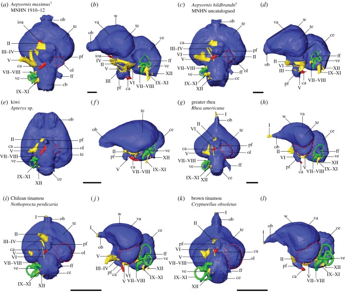
Figure 1.
Digital endocranial reconstructions of (a,b) elephant bird Aepyornis maximus (MNHN F 1910-12), (c,d) elephant bird Aepyornis hildebrandti, (e,f) kiwi, (g,h) greater rhea, (i,j) the open-dwelling Chilean tinamou and (k,l) the forest-dwelling brown tinamou in (a,c,e,g,i,k) ventral and (b,d,f,h,j,l) left lateral views. Optic lobes are highlighted by dashed lines. Colours: blue, brain; green, inner ear; red, vasculature; yellow, cranial nerves. Abbreviations: I, olfactory nerve; II, optic nerve; III, oculomotor nerve; IV, trochlear nerve; V, trigeminal nerve; VI, abducens nerve; VII–VIII, facial and vestibulocochlear nerves; IX–XI, glossopharyngeal, vagus and accessory nerves; XII, hypoglossal nerve; ca, carotid artery; ce, cerebellum; ff, floccular fossa; ioa, internal ophthalmic artery; ob, olfactory bulb; ol, optic lobe (red dashed outline); pf, pituitary fossa; te, telencephalon; va, vallecula; ve, vestibular organs; w, wulst. Scale bars = 1 cm.

