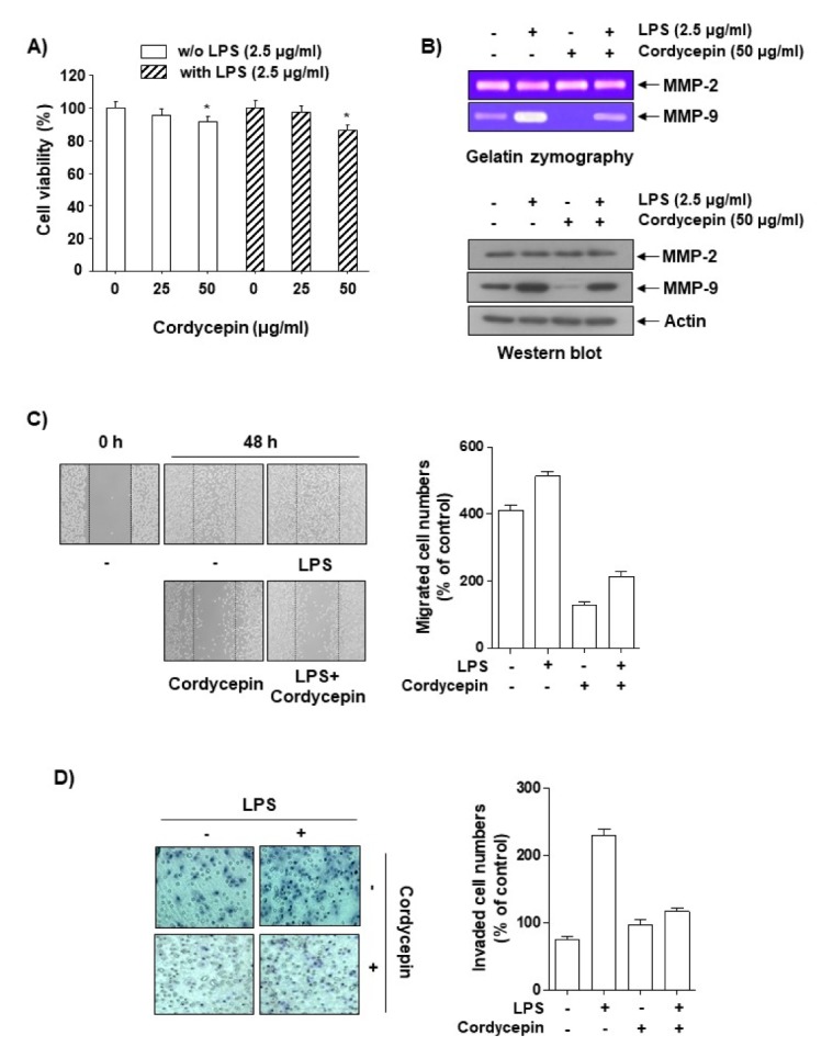
Fig. 1.

Inhibitory effects of cordycepin on the migration and invasion of human colorectal carcinoma HCT116 cells. (A) Cells were incubated with varying concentrations of cordycepin in the absence or presence of LPS for 48 h in serum-free medium, and proliferation was determined using an MTT assay. The data are expressed as the mean ± SD of triplicate experiments. *P < 0.05 compared to control. (B) Cells at 80% confluence were treated with various concentrations of cordycepin in serum-free medium. The conditioned medium was collected after 48 h, and gelatin zymography was performed in triplicate. Representative blots are shown. Equal amounts of cellular proteins were separated on SDS-polyacrylamide gels and transferred to PVDF membranes. The membranes were probed with specific antibodies and visualized using an ECL. Actin was used as an internal control. (C) Cells were scratched with a pipette tip and treated with cordycepin or LPS for 48 h. Migrated cells were imaged using phase-contrast microscopy. (D) The cells were plated onto the apical sides of Matrigel-coated filters in serum-free medium. Medium containing 20% FBS was placed in the basolateral chamber to act as a chemoattractant. After 48 h, cells on the apical side were removed using a Q-tip. The cells on the bottom of the filter were then stained using H&E and photographed.
