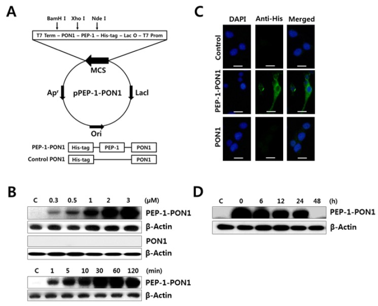
Fig. 1.
Schematic diagram of PEP-1-PON1 expression vector based on the vector pET-15b and the expressed PEP-1-PON1 fusion protein (A). Transduction of PEP-1-PON1 into INS-1 cells (B). For dose-dependent transduction, different concentrations (0.3–3 μM) of PEP-1-PON1 and control PON1 were added to the culture medium for 1 h. For time-dependent transduction, 2 μM PEP-1-PON1 was added to the culture medium for 1–120 min. After incubation, the level of transduced protein was determined by western blotting. Immunofluorescence analysis of the transduced PEP-1-PON1 (C). INS-1 cells treated with 2 μM PEP-1-PON1 or control PON1 were fixed with paraformaldehyde, treated with anti-His-tag primary antibody, and incubated with fluorescein-conjugated secondary antibody. Scale bar, 25 μm. Intracellular stability of the transduced PEP-1-PON1 (D). INS-1 cells pretreated with 2 μM PEP-1-PON1 for 1 h were washed with fresh culture medium and further cultured for 6–48 h.

