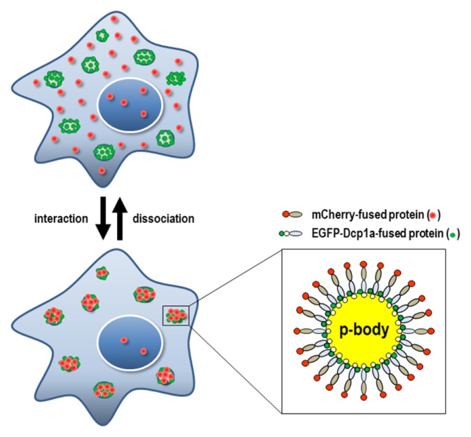
Fig. 1.
Schematic diagram of the SeePPI concept. If mCherry-fused test proteins interact with EGFP-Dcp1a-fused bait proteins, both red and green fluorescent signals will be translocated onto p-bodies at discrete cytoplasmic spots (lower & left figure). Fluorescent fusion proteins co-localized on a p-body were enlarged (lower & right figure). Conversely, when the interaction is disrupted, the red fluorescent signals will be dispersed throughout the cells, leaving a weakened signal on the p-body (upper figure).

