Fig. 2.
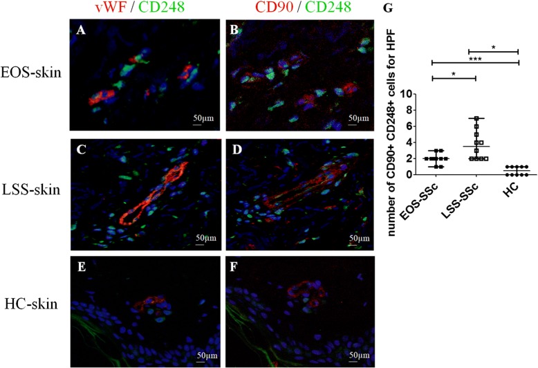
CD248+/CD90+ mesenchymal stem cells (MSCs) surrounding the vessels in systemic sclerosis (SSc) skin. a, b Immunofluorescence staining of ten early-onset subset (EOS) SSc skin samples. Microphotographs show (a) CD248 (green) and von Willebrand factor (vWF) (red) staining and (b) consecutive section stained with CD248 (green) and CD90 (red). c and d Immunofluorescence staining of ten long-standing subset (LSS) SSc skin samples. Microphotographs show (c) CD248 (green) and vWF (red) staining and (d) consecutive section stained with CD248 (green) and CD90 (red). e and f Immunofluorescence staining of ten healthy control subject (HC) skin samples. Microphotographs show (e) CD248 (green) and vWF (red) staining and (f) consecutive section stained with CD248 (green) and CD90 (red). Negative controls were obtained by omitting the primary antibody. Original magnification × 20. g Median number of CD90+/CD248+ cells. The number of CD90+/CD248+ cells is significantly higher in LSS-SSc skin than in EOS-SSc skin. Any dot plot is representative of the median cell count per 5 high-power fields (HPF) (× 40) for each patient. * p = 0.02, *** p = 0.0001

