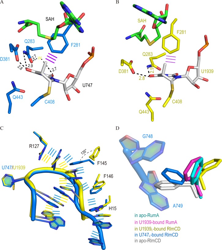
Fig 3. Protein-RNA recognition in complex structures of RlmCD-SAH-U747L and RlmCD-SAH-U1939L.
(A, B) Interaction details between nucleotide of U747 or U1939 and surrounding RlmCD residues. Hydrogen bonding interactions are all indicated as black dashed lines. The yellow “W” represents water molecule and the purple line indicates face-to-edge stacking. (C) Comparison of aromatic stacking interactions in RlmCD-SAH-U747L (marine) and RlmCD-SAH-U1939L (yellow). (D) Side-chain of F145 adopts multiple conformations in apo and RNA-bound structures of RlmCD, while its conformations remain similar in the apo or RNA-bound structure of RumA.

