
Fig. 1.
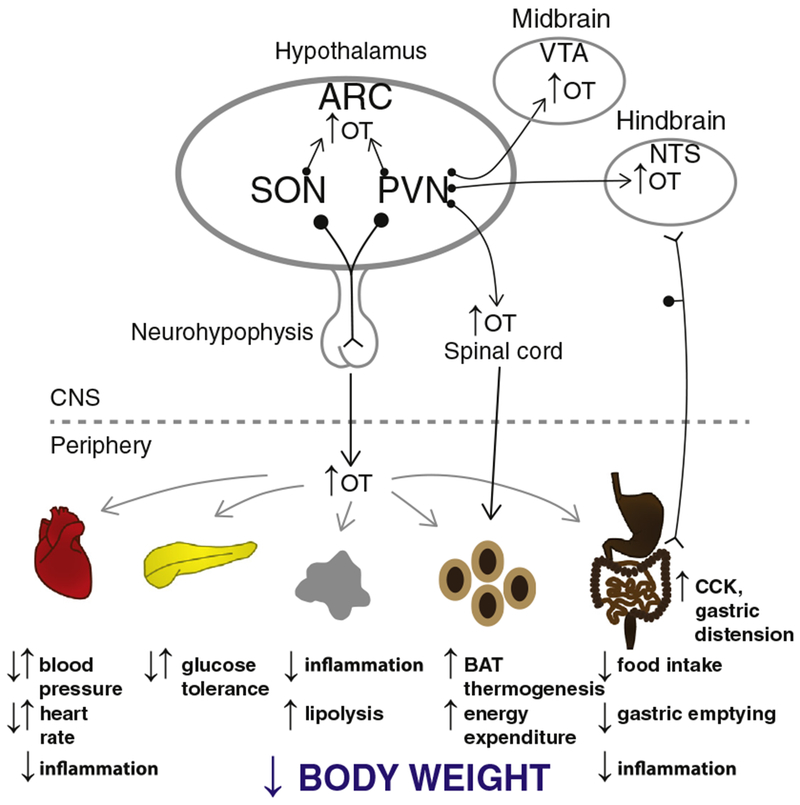
A schematic of circuitry that potentially contributes to the effectiveness of OT on energy homeostasis. OT release within the CNS, spinal cord, and from the neurohypophysis into the circulation (shown in black arrows) may impact metabolic processes (shown in gray) that result in the reduction of bodyweight. Dotted arrow represents implicated pathways from sympathetic preganglionic neurons in the spinal cord to WAT [149,150]. Reported opposing effects denoted by arrows pointed in opposite directions to one another. Abbreviation: ARC, arcuate nucleus; CCK, cholecystokinin; BAT, brown adipose tissue; NTS, nucleus of the solitary tract; PVN, paraventricular nucleus; SON, supraoptic nucleus; and VTA, ventral tegmental area. This figure has been modified from a previous manuscript [Endocrinology Coming full circle: contributions of central and peripheral oxytocin actions to energy balance 154(2):589–96, 2013; http://www.ncbi.nlm.nih.gov/pubmed/23270805].