Fig. 3.
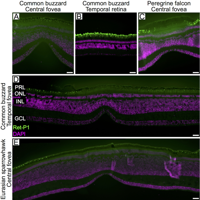
Rhodopsin expression in the rods of the raptor retina. Confocal images of retinal cross-sections labeled with antibodies directed to rhodopsin (Ret-P1; green) of the central fovea (A), the temporal retina (B) and the temporal fovea (D) of the common buzzard, the central fovea of the peregrine falcon (C) and the central fovea of the Eurasian sparrowhawk (E). DAPI used to counter-stain nuclei is shown in purple. For labeling of retinal layers see legend of figure 2. Scale bars: 50 μm.

