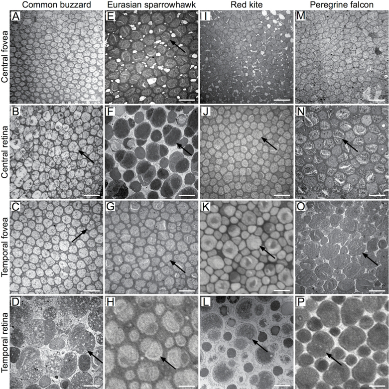
Fig. 4.
Tangential retinal sections through the photoreceptor inner segments of the common buzzard (A, B, C, D), Eurasian sparrowhawk (E, F, G, H), red kite (I, J, K, L) and peregrine falcon (M, N, O, P) retina. TEM (A-G, I, J, L-O) and LM (H, K, P) images of the central fovea (A, E, I, M), the central retina close to the fovea (B, F, J, N), the temporal fovea (C, G, K, O) and the temporal retina (D, H, L, P). Arrows point to the double cones. At the edge of the double cone-free zone of the common buzzard temporal fovea some double cones are also visible (arrow in C). Scale bars: 5 μm.

