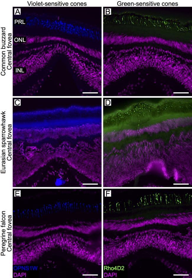
Fig. 6.
Opsin expression in the single cones of the raptor central fovea. Confocal images of retinal cross-sections labeled with antibodies directed to two opsins: SWS1 (violet-sensitive cones; OPN1SW, blue, left column) and Rh2 (green-sensitive cones; Rho4D2, green, right column) of the common buzzard (A, B), Eurasian sparrowhawk (C, D) and peregrine falcon (E, F). DAPI used to counter-stain nuclei is shown in purple. For labeling of retinal layers see legend of figure 2. Scale bars: 50 μm.

