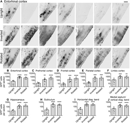
Fig. 2. Inversion and rotation improve the extent and consistency of gene transfer to cerebral cortex and basal forebrain after intrathecal AAV9 infusion.

(A) GFP labeling in medial entorhinal cortex of every animal in the study after intrathecal infusion. GFP expression among rats that remained upright after intrathecal AAV9 infusion was highly variable and minimal in some animals. Gene expression in rats that were inverted or rotated for 2 hours after surgery was both more extensive and more consistent, with robust entorhinal transduction in all 14 animals. Rostral is left in all images. Scale bar, 500 μm. (B to I) Inversion and rotation significantly and substantially enhanced gene transfer to all regions of cortex and basal forebrain. n = 7 rats per cohort. ****P ≤ 0.0001, ***P ≤ 0.001, **P ≤ 0.01: significance of comparison to upright group by one-way analysis of variance (ANOVA) and Tukey’s post test. No significant differences were detected between inverted and rotated rats. Error bars represent SD. Y axes are plotted on a logarithmic scale.
