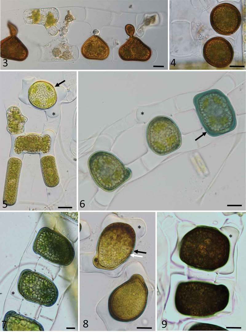
Figs 3–9.

Light microscopy images of zygospores of Zygnema cf. calosporum (Figs 3, 4) and Zygnemopsis lamellata (Figs 5–9) collected in Svalbard. Fig. 3. Conjugating filaments, with partially developed zygospores; Fig. 4. mature zygospores; Fig. 5. conjugating stage (arrow) and fully fused zygospore (arrow) with massive appendages (asterisk); Fig. 6. zygospores with blue mesospore (black arrow) at different stages of development; Fig. 7. zygospores still in conjugating filaments, positioned in the middle, with appendages (asterisk); Fig. 8. zygospores with a two-layered mesospore clearly visible, comprising an outer layer in blue (black arrow), and an inner layer scrobiculate (white arrow), with appendages marked by an asterisk; Fig. 9. dark appearance of fully developed zygospores, showing appendages of the exospore (asterisk); Scale bars: Figs 3–9 = 20 µm.
