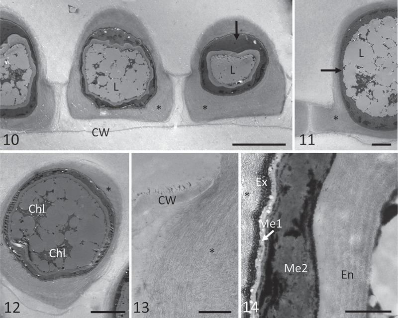
Figs 10–14.

Transmission electron micrographs of zygospores of Zygnemopsis lamellata collected in the field. Fig. 10. Several zygospores still in the original filaments, showing an electron-dense inner mesospore layer (arrow), and exospore surrounded by pectic-cellulosic appendages (‘wings’) marked with an asterisk; Fig. 11. electron-dense mesospore layer (arrow), with lipid bodies inside the zygospores, and appendages marked by an asterisk; Fig. 12. chloroplasts in zygospores; Fig. 13. pectic-cellulosic appendage within the gametangium; Fig. 14. exospore (Ex), two-layered mesospore (Me1 = electron-translucent, Me2 = electron-dense and irregular) and highly sculptured endospore (En). Abbreviations are Chl = chloroplast, CW = cell wall of the mother cell, Ex = exospore, L = lipids, Me1 = outer mesospore layer, Me2 = inner mesospore layer, En = endospore. Scale bars: Fig. 1 = 20 µm; Figs 11–12 = 10 µm; Fig. 13 = 2 µm; Fig. 14 = 1 µm.
