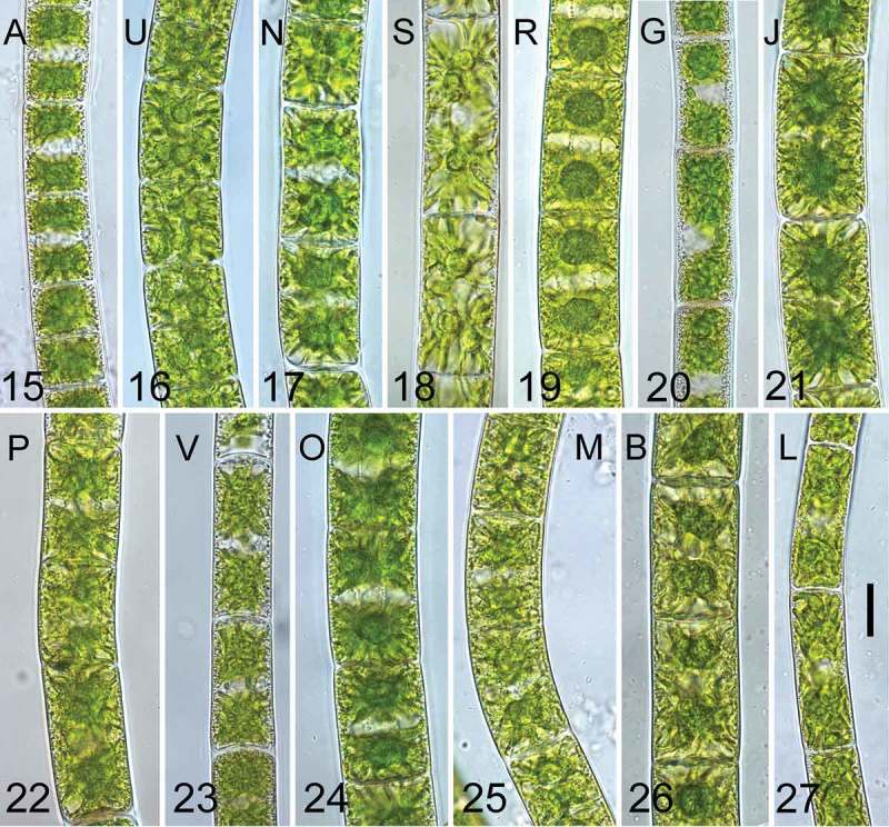
Figs 15–27.

Young vegetative cells (3 weeks after transfer to fresh medium) of the investigated genotypes. Fig. 15. Genotype A; Fig. 16., U; Fig. 17. N; Fig. 18. S; Fig. 19. R; Fig. 20. G; Fig. 21. J; Fig. 22. P; Fig. 23. V; Fig. 24. O; Fig. 25. M; Fig. 26. B; Fig. 27. L. Scale bar = 20 µm in all images. Genotypes are ordered according to phylogeny described in Fig. 2.
