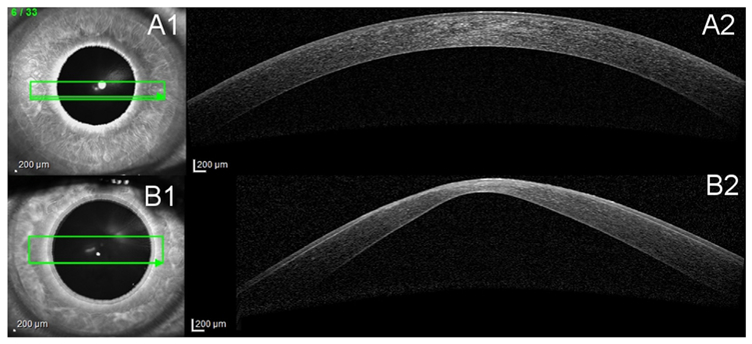
Fig. 3.

Anterior cross-section Optical Coherence Tomography (OCT). A1 and B1 show the scan location on the cornea. A2 and B2 show the cross-section view of the cornea. A: Healthy cornea. B: Severe KC cornea, characteristic protrusion and thinning as well as scarring at the top of the cone.
