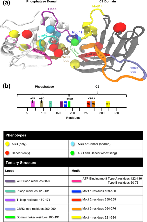
Figure 2.
Mapping of ASD- vs. cancer-associated mutations on PTEN three-dimensional structure. (a) Individual spheres correspond to 17 germline missense PTEN mutations represented by ASD only (yellow), cancer only (red), mutations shared across both phenotypes (cyan), one mutation with coexisting ASD and cancer (green). Mutations occur in both the phosphatase domain (white) and C2 domain (grey) of WT PTEN structure, (b) Domain structure of PTEN protein.

