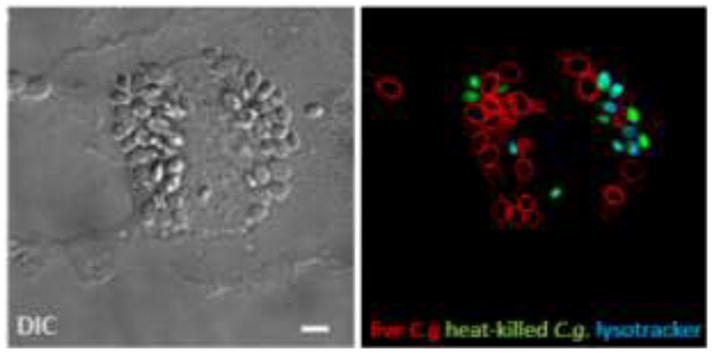
Figure 1.

Phagocytosed C. glabrata by a macrophage. Live (red) and heat-killed C. glabrata (green) were incubated with a mouse primary bone-marrow derived macrophage. Lysotracker (blue) delineates the acidified compartments containing only dead yeast, whereas live C. glabrata evades phagosomal acidification remaining in a neutral compartment. Bar = 5μm.
