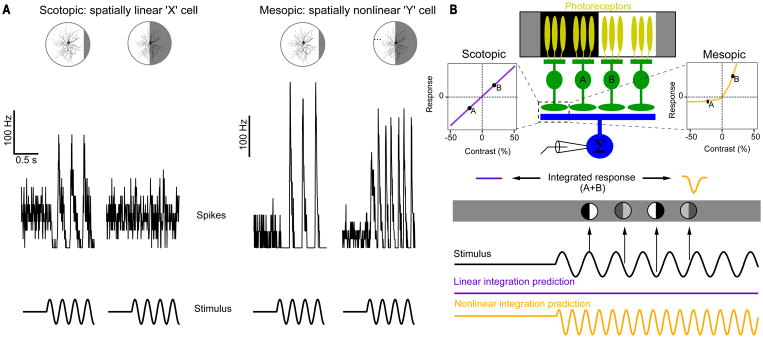
Figure 8. Linear verses nonlinear spatial integration in RGCs can depend on luminance.
(A) Example of the same ON-alpha responding to a contrast-reversing grating in scotopic and mesopic luminance. This is the same stimulus originally used to classify linear (X) vs. nonlinear (Y) RGCs (Enroth-Cugell and Robson, 1966). (B) A schematic of how a change in rectification at bipolar cell output synapses can account for a change in spatial integration in a RGC. Figure adapted from (William N. Grimes et al., 2014).

