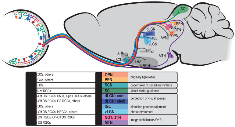
Figure 9. A subset of retinorecipient areas of the brain.
Well studied brain regions are in color and listed with their known RGC inputs and their behavioral function. Less well understood regions are shown shaded in gray.
Abbreviations: AHN: Anterior Hypothalamic Nucleus, APN: Anterior Pretectal Nucleus, IGL: Intergeniculate Leaflet, LGN: Lateral Geniculate Nucleus, LHA: Lateral Hypothalamic Area, MTN: Medial Terminal Nucleus, NOT/DTN: Nucleus of the Optic Tract/Dorsal Tegmental Nucleus, OPN: Olivary Pretectal Nucleus, PPN: Pedunculopontine Nucleus, RCH: Retrochiasmatic Area, SC: Superior Colliculus, SCN: Suprachiasmatic Nucleus.

