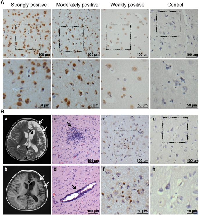
Fig. 1.
HHV6 expression in brain tissues of patients with RE and controls, as well as characteristics of magnetic resonance imaging (MRI) and histopathological changes in the RE case. A Representative images of strong, moderate, and weak positive staining and negative staining for the HHV6 antigen under low (scale bar = 100 µm) and high (scale bar = 50 µm) magnification. Neuron-like cells were stained. B Atrophy of the left hemisphere cortex (white arrows) and widening of the caudate nucleus (black arrow) were observed in the patient by T2 (B-a) and FLAIR (B-b) images. HE staining shows microglial nodule formation (B-c), lymphocyte infiltration, and perivascular cuff (B-d) in the temporal lobe cortex of the brain. Activation of CD8+ T cells was detected in the RE (B-e) and (B-f) but not in the brain tissue from the trauma patient (B-g) and (B-h) by IHC under low (scale bar = 100 µm) and high (scale bar = 50 µm) magnification.

