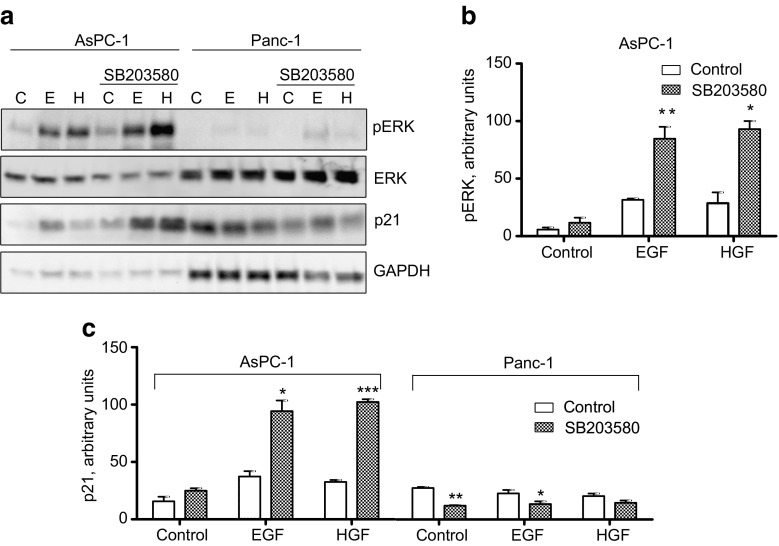
Fig. 5.
Effect of p38 inhibition on the expression of p21. AsPC-1 and Panc-1 cells were stimulated by EGF (10 nM) or HGF (1 nM) for 24 h with or without SB203580 (10 μM) pretreatment for 30 min. (a) Immunoblots showing p21 expression and ERK phosphorylation. (b) Densitometric quantification of ERK phosphorylation in AsPC-1 cells based on three independent experiments. The level of pERK was normalized to total ERK. (c) Densitometric quantification of p21 expression in AsPC-1 and Panc-1 cells based on three independent experiments. p21 expression was normalized to the level of GAPDH. The data represent the mean ± SEM of three independent experiments. * indicates p ≤ 0.05, **indicates p ≤ 0.01 and *** indicates p ≤ 0.001 versus without SB203580

