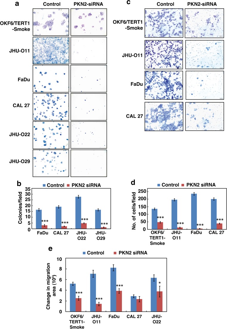
Fig. 5. Inhibition of PKN2 decreases the invasive and migratory ability of smoke exposed cells and HNSCC cells exposed to cigarette smoke.
a–b Colony formation assays were carried out using OKF6/TERT1-Smoke, JHU-O11, FaDu, CAL 27, JHU-O22 and JHU-O29 cells following siRNA-mediated silencing of PKN2 or control siRNA (scrambled siRNA), A graphical representation of the colony forming ability of the indicated cells upon PKN2 silencing (*p ≤ 0.05; **p ≤ 0.01; ***p ≤ 0.001). Colonies were visualized at 3× magnification. c–d Invasion assays were carried out using OKF6/TERT1-Smoke, JHU-O11, FaDu and CAL 27 cells. Cells were transfected with either control (Scrambled) or PKN2 siRNA and invaded cells were photographed at 10× magnification. A graphical representation of the invasive ability of the cells upon PKN2 silencing (*p ≤ 0.05; **p ≤ 0.01; ***p ≤ 0.001) (e) Wound migration assays were carried out using OKF6/TERT1-Smoke and HNSCC cells with or without PKN2 SiRNA. Distance between migrated cells were calculated between 0 and 10 h and represented as bar graph. (*p ≤ 0.05; **p ≤ 0.01; ***p ≤ 0.001)

