Fig. 4.
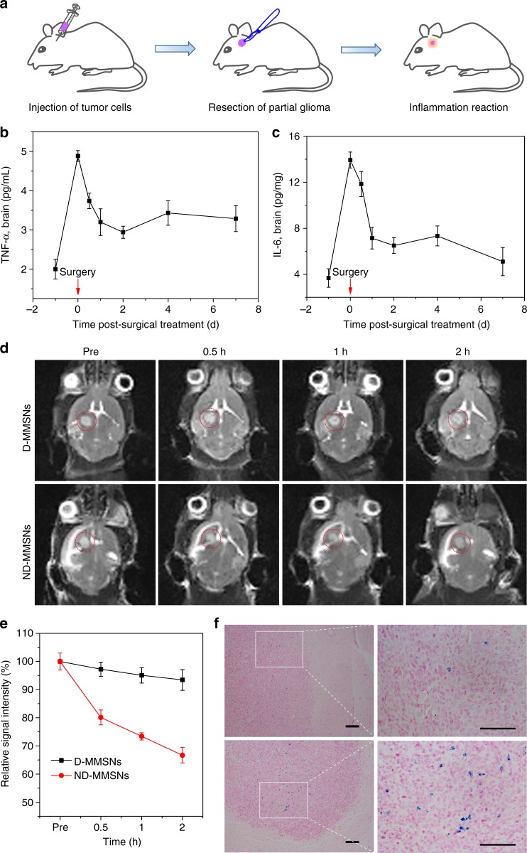
In vivo T2-weighted MR imaging. a Schematic representation of in vivo inflammation reaction after resecting partial glioma. Evaluation of the inflammation cytokines TNF-α (b) and IL-6 (c) levels in the brain of the U87-bearing mice after surgical treatment for 7 d. d In vivo T2-weighted MR images of postsurgical glioma-bearing mice before and after intravenous injection of D-MMSNs and ND-MMSNs. The red circles point to the tumor site. e Relative signal intensities of postsurgical glioma as a function of time. f Histological analysis of brain tissues by Prussian blue staining. Scale bar: 50 μm. Mean values and error bars are defined as mean and s.d., respectively (n = 3)

