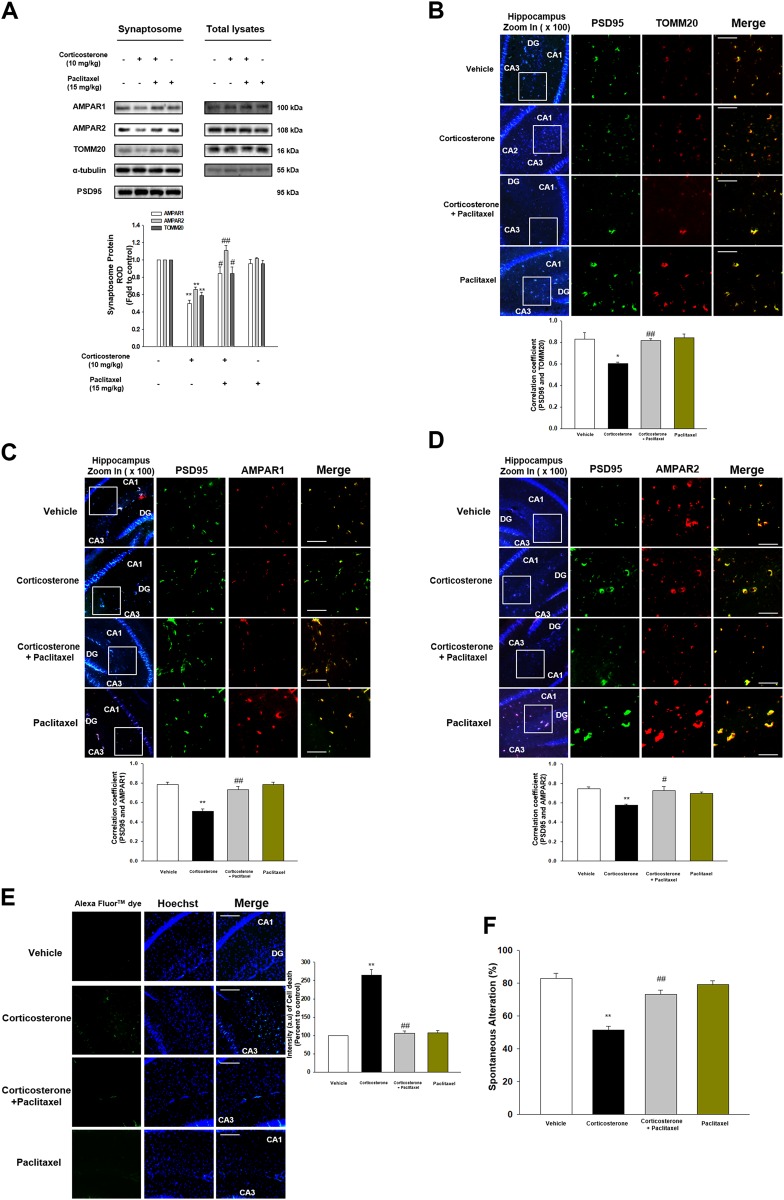
Fig. 6. Corticosterone-induced memory impairment was attenuated by paclitaxel.
a The synaptosome of hippocampus from mice exposed to vehicle, corticosterone (10 mg/kg), corticosterone with paclitaxel (15 mg/kg), and paclitaxel alone was isolated. AMPAR1, AMPAR2, TOMM20, α-tubulin, and PSD95 were detected. PSD95 was used as a loading control of synaptosome. ** indicates p < 0.01 vs. vehicle. #, ## indicates p < 0.05, p < 0.01 vs. corticosterone treatment group, respectively. n = 6. b–d The mice were exposed to vehicle, corticosterone (10 mg/kg), corticosterone with paclitaxel (15 mg/kg), or paclitaxel. Slide samples for IHC were immunostained with PSD95 (green), TOMM20 or AMPAR1/2 (red), and DAPI (blue). *, ** indicates p < 0.05, p < 0.01 vs. vehicle, respectively. #, ## indicates p < 0.05, p < 0.01 vs. corticosterone alone, respectively. Scale bars, 200 μm (magnification, ×200). n = 5. e TUNEL assay was performed using slide samples of hippocampus from mice with vehicle, corticosterone (10 mg/kg), corticosterone with paclitaxel (15 mg/kg), or paclitaxel. The intensity of green fluorescence indicates the amount of neuronal cell death. ** indicates p < 0.01 vs. vehicle and ## indicates p < 0.01 vs. corticosterone alone. Scale bars, 200 μm (magnification, ×200). n = 5. f The mice exposed to vehicle, corticosterone (10 mg/kg), corticosterone with paclitaxel (15 mg/kg), or paclitaxel were subjected to Y-maze test to evaluate memory function. ** indicates p < 0.01 vs. vehicle and ## indicates p < 0.01 vs. corticosterone alone. n = 6

