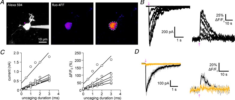
Figure 2. One‐photon uncaging of DPNB‐ABT594 on medial habenula neurons with violet light.

A, two‐photon fluorescence maximum projection image of a patch‐clamped medial habenula (MHb) neuron. Left: morphology of a MHb neuron filled with Alexa 594 with the position of uncaging labelled (violet). Middle and right: pseudo‐colour images showing the increase in fluorescence of fluo‐4FF (right compared to baseline, middle) caused by DPNB‐ABT594 (20 μM, bath‐applied) one‐photon uncaging. Dashes indicate region of interest used for analysis. B, amplitudes of inward current (left) and fluo‐4FF fluorescence (right) in response to increased duration of irradiation (410 nm, 1 mW, 0.5–3.0 ms). C, uncaging‐evoked currents and Ca2+ signals increase linearly with uncaging duration. Inward current amplitudes (left) and fluo‐4FF fluorescence amplitudes (right) plotted against duration of irradiation for each cell (n = 5 cells; 410 nm, 1 mW). Linear regression lines were fitted. D, currents and fluo‐4FF fluorescent signals were blocked (yellow traces) by the nAChR antagonists DHβE (3 μM) and mecamylamine (3 μM). Black/dark yellow traces are averages of three consecutive trials (grey/light yellow). [Color figure can be viewed at http://wileyonlinelibrary.com]
