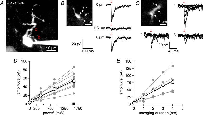
Figure 5. Two‐photon uncaging of DPNB‐ABT594 on medial habenula neurons.

A, two‐photon fluorescence image of a patch‐clamped medial habenula (MHb) neuron filled with Alexa 594 (maximum projection of 25 μm). Regions selected for two‐photon uncaging at 720 nm of DPNB‐ABT594 (400 μM, locally applied) are marked by the letters b and c and are shown in B and C at higher resolution. B, current response (right, top) caused two‐photon uncaging (2 ms, 30 mW) on the dendrite (top left, zoomed image of dendrite b in A). Current response (right, middle) from uncaging at 1.5 μm away from the dendrite. Redirecting the laser to the dendrite restored the current response (right, bottom). Current traces are averages of 3–4 trials. C, current recordings in response to increasing power (0–40 mW) with constant duration (2 ms) at locations 1–3 (top left, zoomed image of dendrite c in A). D, two‐photon uncaging‐evoked currents increased quadratically with power (n = 9 cells with 3 power curves each; 2 ms, 0–40 mW). Uncaging on cells without DPNB‐ABT594 (×10, 40 mW, 2 ms, at 5 Hz) evoked no current (square, n = 4). E, two‐photon uncaging‐evoked currents showed a linear dependence on photolysis duration (0–4 ms) at constant power (20 mW, n = 4 cells with 3 power curves each). Linear regression lines in D and E were fitted for each cell (grey points and lines) and the average across cells (white points and black lines). [Color figure can be viewed at http://wileyonlinelibrary.com]
