
Fig. 1.
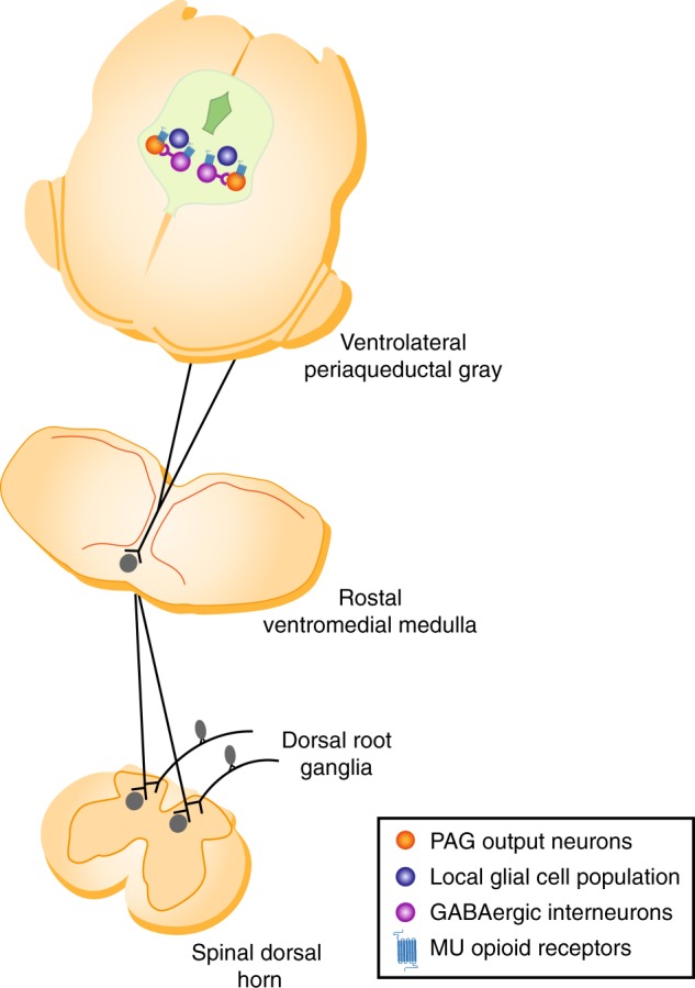
A schematic of the descending inhibitory pathway for pain modulation illustrating the projections from the caudal ventrolateral column of the midbrain periaqueductal gray to the rostral ventromedial medulla in the brainstem and the dorsal horn of the spinal cord at the level of incoming stimulation from sensory neurons of the dorsal root ganglia. Also indicated are local GABAergic interneurons (purple) and PAG–RVM output neurons (orange) and a local glial cell population (blue) that also signals to the PAG–RVM output neurons. Mu opioid receptors expressed on local GABAergic interneurons and PAG–RVM output neurons are also indicated