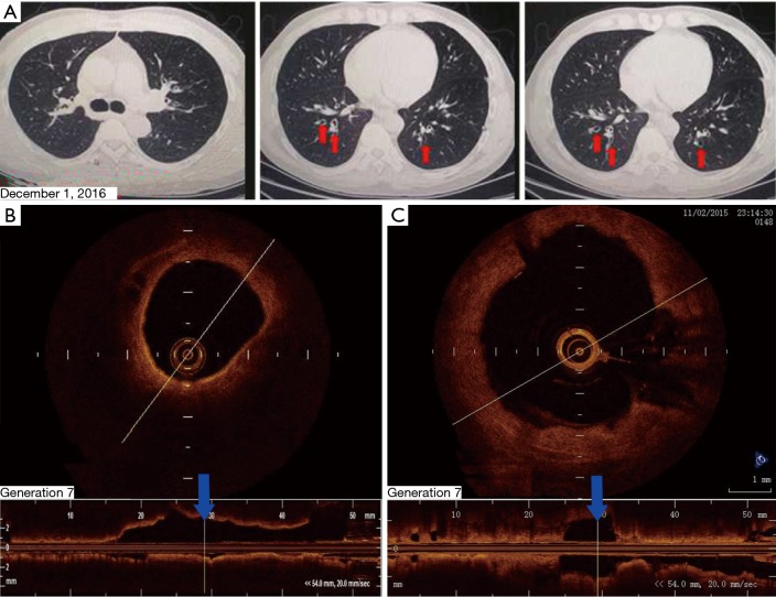
Figure 3.
Bronchiectasis of the patient were evaluated again by chest computed tomography (CT) and optical coherence tomography (OCT) as following: (A) chest CT scan after bronchial thermoplasty (BT) on December 1, 2016. The red arrows indicate mild bilateral bronchiectasis. R indicates the right side; (B) on OCT performed on February 24, 2017, the 4th- to 5th-generation bronchi were mildly dilated; (C) the OCT image recorded on November 2, 2015 shows that the cross section of the 6th- to 7th-generation bronchi in the posterior basal segmental bronchus of the left lower lobe to be significantly dilated. The distal bronchus was narrow and obstructed but lacked sputum bolt.

