
Figure 2.
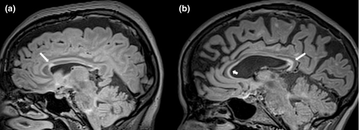
Parasagittal FLAIR images showing the appearance of Corpus Callosum lesions in a patient with Fabry Disease (a) and in one with Multiple Sclerosis (b). In the MS patient is possible to better appreciate, compared to FD, the typical appearance of the calloso‐septal lesions, which are defined as narrow hyperintense bands along the undersurface of the corpus callosum itself