Figure 2.
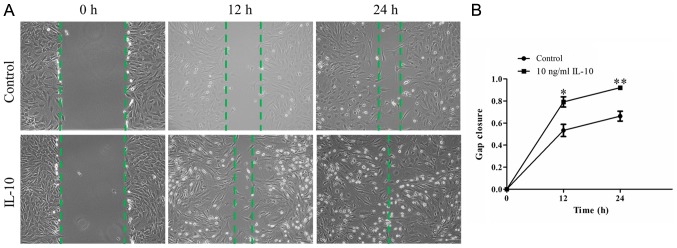
IL-10 promotes TDSC migration (A) Representative time-lapse migration images of control and IL-10-treated cells from the wound healing assay. Images were acquired 0, 12 and 24 h after scratching. Original magnification, ×100. (B) The migration rate was measured by quantifying the total area of the entire strip lacking cells. The relative migration rate of recovered area at 12 and 24 h was calculated from three independent experiments. Data are expressed as the mean ± standard error of the mean. *P<0.05, **P<0.01 vs. control cells. IL-10, interleukin-10; TDSCs, tendon-derived stem cells.

