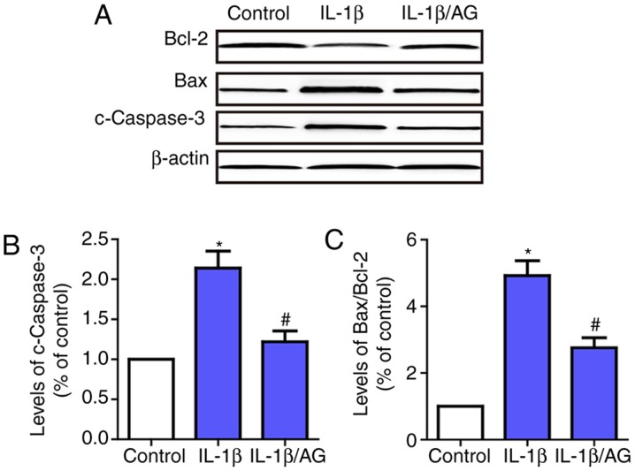
Figure 2.
Effects of AG on IL-1β-induced apoptosis of NP cells. Cells were stimulated by IL-1β with or without AG treatment. (A) Western blot analysis was used to detect the levels of c-caspase-3 and Bax/Bcl-2. (B) Quantification of c-caspase levels. (C) Quantification of the Bax/Bcl-2 ratio. β-actin was used as an internal control. The values are presented as the mean ± standard deviation of 3 independent experiments. *P<0.05 vs. control group; #P<0.05 vs. IL-1β group. AG, andrographolide; NP, nucleus pulposus; IL-1β, interleukin 1β; c-caspase-3, cleaved caspase-3; Bcl-2, B-cell lymphoma 2; Bax, Bcl-2-associated X protein.

