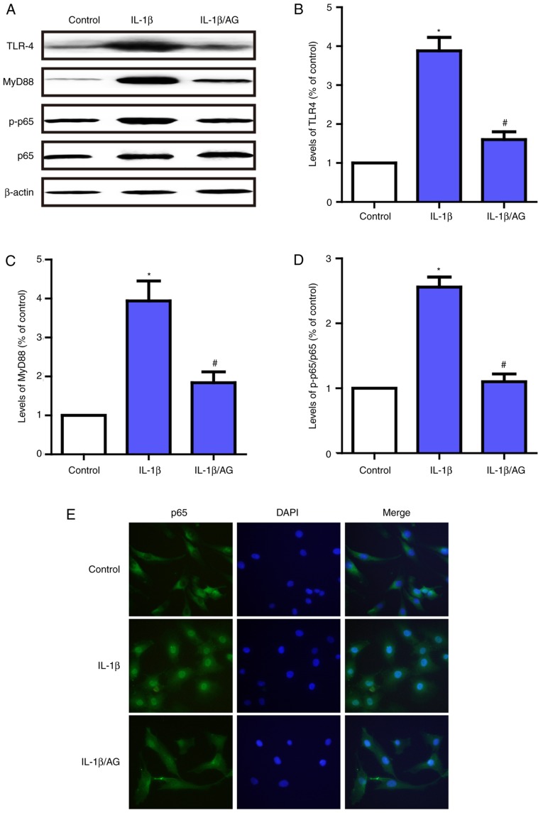
Figure 5.
Effect of AG on IL-1β-induced activation of the TLR4/MyD88/NF-κB signaling pathway in human NP cells. Cells were stimulated by IL-1β with or without AG treatment. (A) The levels of TLR-4, MyD88, p65 and p-p65 were determined by western blot analysis. (B) Quantification of TLF4 levels. (C) Quantification of MyD88 levels. (D) Quantification of p65/p-p65 ratio. (E) p65 expression was also measured by immunofluorescence (magnification, ×400). The values are presented as the mean ± standard deviation of 3 independent experiments. *P<0.05 vs. control group; #P<0.05 vs. IL-1β group. AG, andrographolide; IL-1β, interleukin 1β; TLR-4, toll-like receptor 4; MyD88, myeloid differentiation primary response protein MyD88; p65, Transcription factor p65; p, phosphorylated.

