Figure 1.
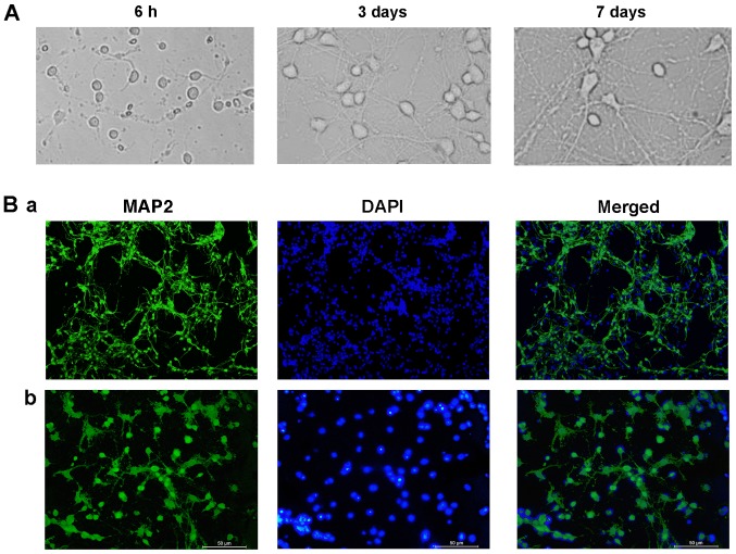
Morphology and purity of hippocampal neurons cultured in vitro. (A) Morphology of hippocampal neurons at various time points. (B) Immunofluorescence in 7 DIV hippocampal neurons. Neurons were stained with MAP2 and DAPI, and images are merged. Neurons were observed using an inversion fluorescence microscope and representative images at (Ba) magnification ×40 and (Bb) magnification ×200 are presented. MAP2, microtubule-associated protein 2.

