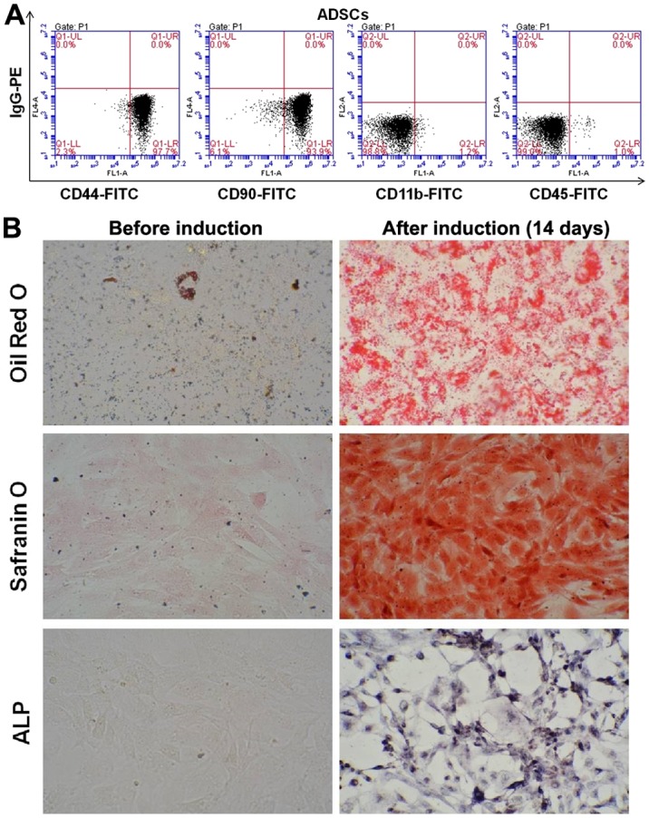
Figure 1.

The identification of the 3rd generation ADSCs using flow cytometry. (A) ADSCs were positive for the cell surface markers CD44 (97.7%) and CD90 (93.9%) but negative for CD11b (1.1%) and CD45 (1.0%) (n=3). (B) The multipotentiality of ADSCs cells. Oil Red O staining of the ADSCs after 2 weeks of culture demonstrated numerous intracellular lipid droplets. Safranin O and alkaline phosphatase staining was positive. Magnification, ×200. ADSCs, adipose-derived stem cells.
