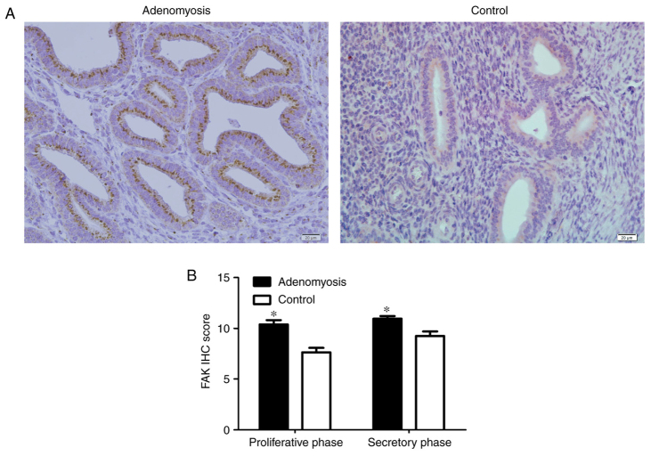
Figure 1.
IHC staining of FAK in human endometrium. (A) IHC analysis of FAK in eutopic endometrium from adenomyosis and control tissues. Dark yellow to brown presents positive staining. Magnification, ×100. (B) Scoring analysis of FAK IHC staining in different phases. *P<0.05 vs. Control. FAK, focal adhesion kinase; IHC, immunohistochemical.

