
Figure 2.
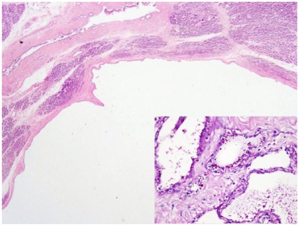
Low-power view of unilocular variant exhibiting a single large cystic locule lined by single layer of cells. Inset: PAS special stain highlighting intracytoplasmic glycogen granules.
Official websites use .gov
A
.gov website belongs to an official
government organization in the United States.
Secure .gov websites use HTTPS
A lock (
) or https:// means you've safely
connected to the .gov website. Share sensitive
information only on official, secure websites.
Low-power view of unilocular variant exhibiting a single large cystic locule lined by single layer of cells. Inset: PAS special stain highlighting intracytoplasmic glycogen granules.