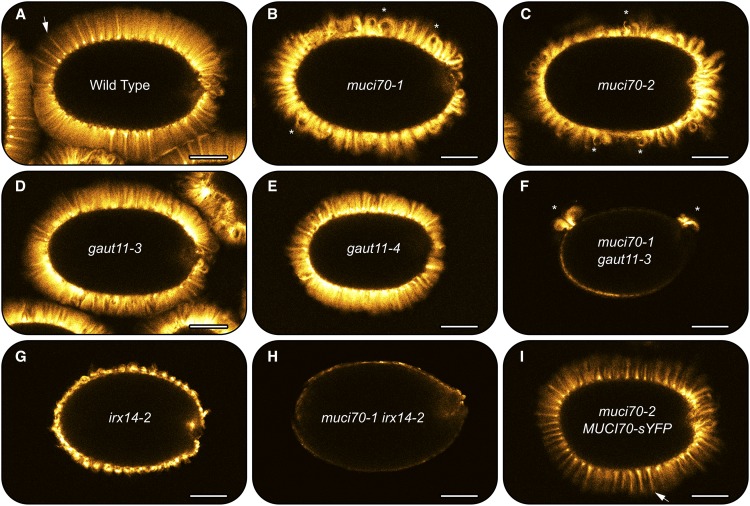
Figure 6.
S4B staining of cellulose in mucilage capsules of seeds hydrated in water. Single optical sections of fluorescent signals were detected with a confocal microscope. Arrows show well-defined cellulosic rays (A and I). Asterisks indicate short, curly rays observed in mutants with muci70 insertions. No straight rays are observed in F to H. Bars = 150 µm.

