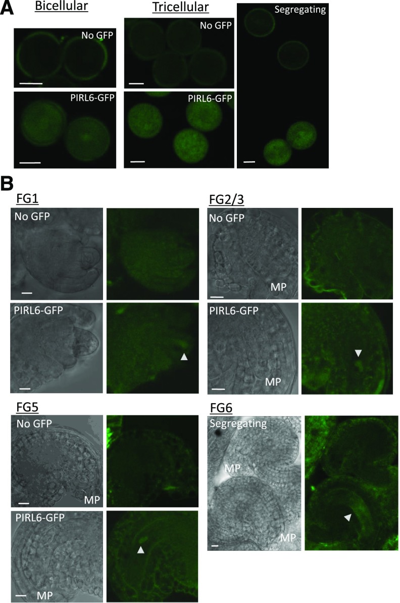
Figure 2.
Expression of PIRL6-GFP in both male and female gametophytes. Gametophytes segregating for a full-length PIRL6-GFP fusion construct were viewed at the indicated developmental stages by confocal and differential interference contrast (DIC) microscopy. Plants were hemizygous for the reporter construct, producing pollen and embryo sacs that segregated 1:1 for the PIRL6-GFP construct; for each developmental stage shown above, pollen and ovules were from the same anthers and ovaries, respectively. A, Expression in bicellular and tricellular stage pollen; the right-most image is a single image illustrating the meiotic segregation of the reporter construct in pollen from a hemizygous anther. B, Expression during female gametophyte development. Triangles mark locations of PIRL-GFP within the embryo sac. Female gametophyte (FG) developmental stages were defined by Christensen et al. (1998). To provide a reference point, the micropylar end of each ovule is labeled on the DIC panels (MP). The FG6 stage image is a single image illustrating the segregation of the reporter construct in adjacent ovules. Bars = 10 μm.

