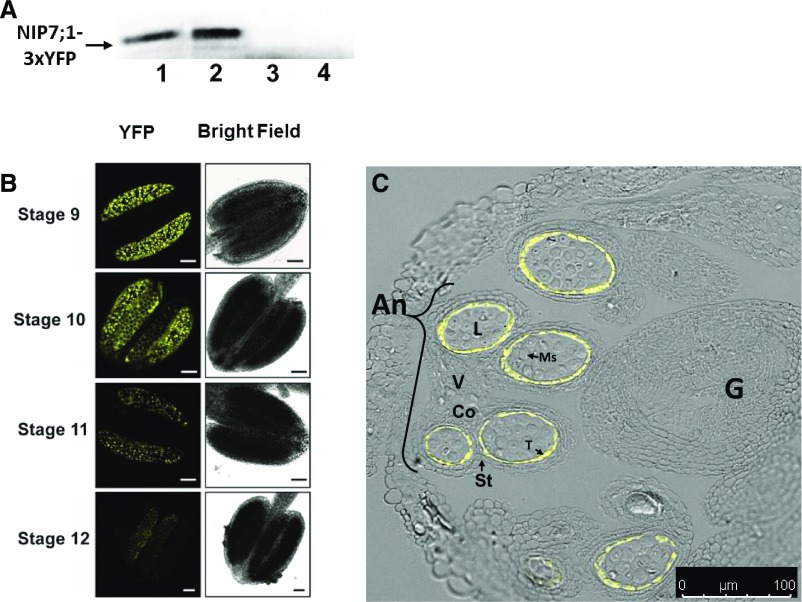
Figure 4.
Localization of AtNIP7;1 protein in developing Arabidopsis anthers. A, Western-blot analysis of AtNIP7;1 from extracts (10 μg of extract protein per lane) of dissected Arabidopsis flower buds at the following stages: lane 1, stages 9 and 10; lane 2, stages 10 and 11; lane 3, stage 12; and lane 4, stages 13 and 14. The arrow indicates the position (100 kD) expected for the NIP7;1-3xYFP protein product. B, Confocal fluorescence micrographs of live anthers of NIP7;1:3xYFP recombineering plants at the indicated floral stages. Bars = 50 μm. C, Confocal fluorescence micrograph of an anther section from a stage 10 flower (anther stage 9) of NIP7;1:3xYFP recombineering plants superimposed on a differential interference contrast image of an anther with various cell types indicated. An, Anther; Co, connective; G, gynoecium; L, locule; Ms, pollen microspores; St, stomium; T, tapetum; V, vascular region. Bar = 50 μm.

