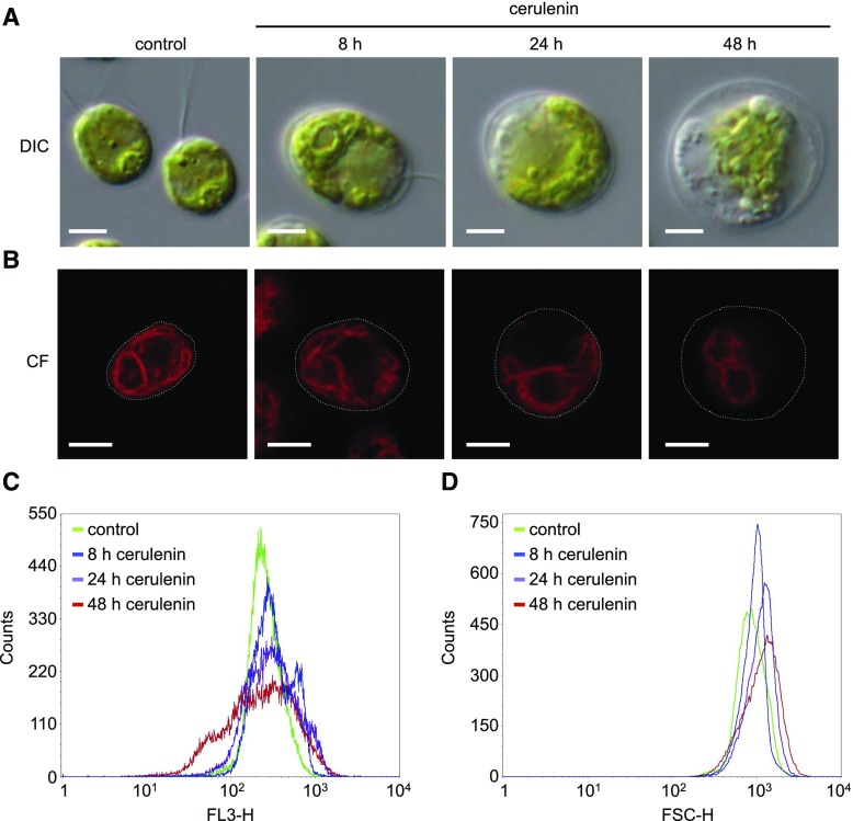
Figure 4.
Effects of cerulenin on Chlamydomonas cell morphology. A, DIC microscopy images of log-phase Chlamydomonas cells treated with 10 μm cerulenin for 8, 24, and 48 h. Untreated cells were used as a control. Bars = 5 μm. B, Chlorophyll fluorescence (CF) from individual cells treated with 10 µm cerulenin for 8, 24, and 48 h was visualized by confocal microscopy. Bars = 5 μm. C and D, Chlorophyll fluorescence (C) and size (D) of 5 × 10⁴ cells treated with 10 µm cerulenin for 8, 24, and 48 h were analyzed and quantified by flow cytometry. FL3-H, Fluorescence (red channel); FSC-H, Forward-scattered light (size).

