Figure 6.
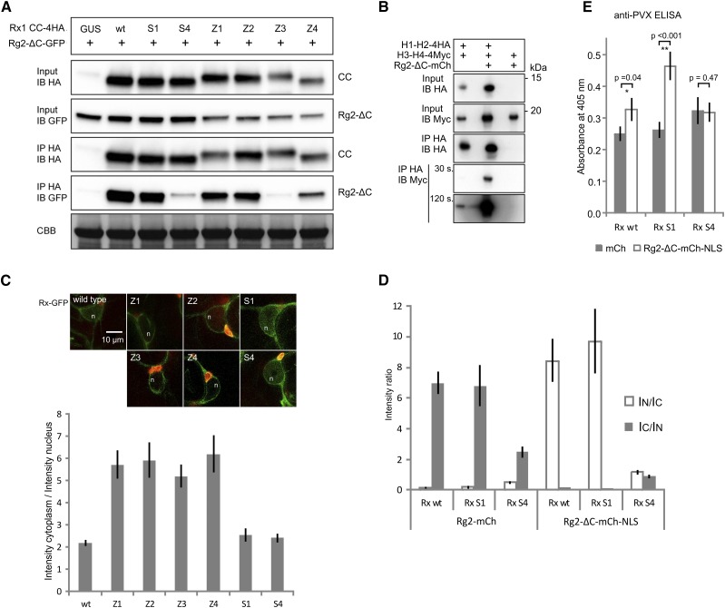
Effects of the mutations in the CC on the interaction with RanGAP2 and the subcellular localization of Rx1. A, Coimmunoprecipitation of HA-tagged versions of the Rx1 CC variants Z1 to Z4, S1, S4, and the wild type (wt) with the N-terminal WPP domain of RanGAP2 (Rg2-ΔC-GFP). Equal loading of the input material is shown by the Coomassie Brilliant Blue-stained Rubisco (CBB). B, Effects of the presence of the RanGAP2 WPP domain on the interaction between H1-H2 and H3-H4. Anti-HA immunoprecipitation of H1-H2-4HA coexpressed with H3-H4-4Myc in the presence or absence of mCherry-tagged RanGAP2 WPP domain (Rg2-ΔC-mCh) is shown. Two exposures (30 and 120 s) are shown for the anti-Myc immunoblot with the results of the anti-HA immunoprecipitation to show the two bands of different intensity. C, Full-length Rx1 constructs (wild type, Z1–Z4, S1, and S4) with a C-terminal GFP fusion were imaged using confocal microscopy after 2 d of expression in N. benthamiana leaves. The images show nuclei (n) and surrounding cytoplasm in representative cells. Chlorophyll autofluorescence is shown in red. Bar = 10 μm for all images. The ratio of GFP fluorescence intensity in the cytoplasm and nucleus was determined in seven to 12 cells for each construct. The graph shows the average cytoplasmic intensity/nuclear intensity ratio (IC/IN). The error bars represent the se. Higher values indicate a more cytoplasmic localization profile. D, Coexpression of Rx-GFP variants with RanGAP2 constructs to test the effect on the localization of Rx. Rx-GFP (wild type, S1, and S4) was coexpressed with either full-length RanGAP2-mCherry (Rg2-mCh) or with Rg2-ΔC-mCh-NLS, a construct in which the RanGAP2 WPP domain was tagged to a nuclear localization signal. The localization of wild-type Rx is affected by the coexpression of these constructs: RanGAP2 sequesters it in the cytoplasm, and Rg2-ΔC-mCh-NLS targets it to the nucleus. The GFP intensities from the Rx-GFP constructs were determined for the nucleus and cytoplasm, and the average ratios of the intensities are plotted (n = 9; error bars denote the se). E, PVX resistance assay. Full-length Rx (wild type, S1, and S4) was coexpressed with either mCherry as a control or with Rg2-ΔC-mCh-NLS and an avirulent PVX amplicon. Previously, we showed that targeting full-length Rx to the nucleus led to a partial loss of resistance (Slootweg et al., 2010). The level of virus after 5 d was determined by an anti-PVX CP ELISA. Error bars represent the se (n = 9). Student’s t test was used to determine if coexpression of Rg2-ΔC-mCh-NLS resulted in a significantly higher virus level than coexpression with free mCherry (*, P < 0.5 and **, P < 0.05).

