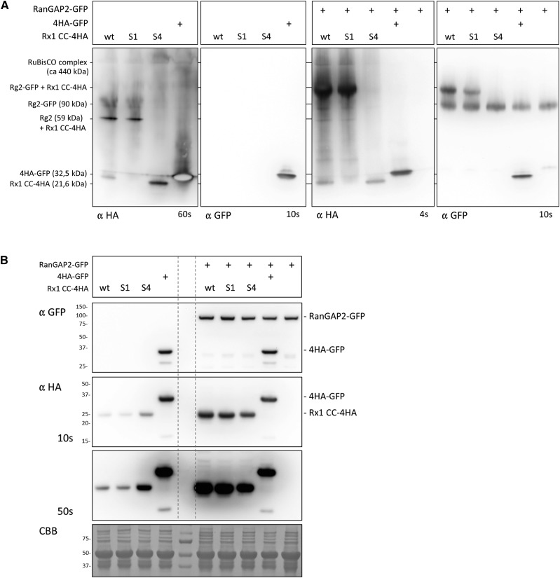
Figure 7.
Blue Native gel analysis of the complex formed by the Rx1 CC and RanGAP2 in the cell. A, Blue Native gel analysis of Rx1 CC constructs (wild type [wt], S1, and S4) coexpressed with RanGAP2-GFP (right two images) or expressed alone (left two images). HA-tagged GFP (4HA-GFP) was included as a control and was detected with the anti-HA and anti-GFP antibody. The mass given for the RanGAP2, Rx1-CC, and GFP constructs represents the mass of a monomer, and for Rubisco the approximate mass of the complex is given. The behavior of the proteins on this gel is determined by the mass of the complex they are part of and by their shape. The blots were aligned to each other using the Rubisco complex and the 4HA-GFP, which is present on each immunoblot. B, SDS-PAGE analysis of the samples used in A demonstrating that the banding patterns on the Native Blue blot are not due to protein degradation or modifications. RanGAP2-GFP and the CC-4HA constructs run as single bands on SDS-PAGE. A Coomassie Brilliant Blue (CBB)-stained blot is included as a control for equal loading. The dashed vertical lines indicate the positions of the marker lane on the immunoblots.

