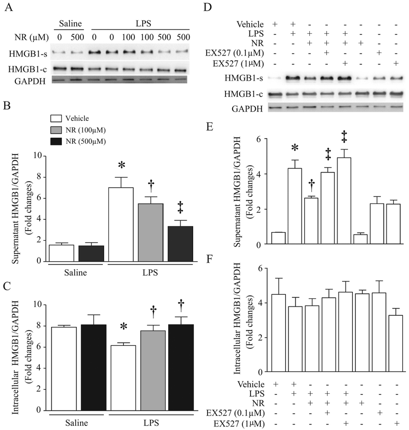
Fig. 6.
Effects of NR on LPS-induced HMGB1 release in Macrophages. (A–C) Cultured RAW264.7 cells were pretreated with NR (100 μM, 500 μM) or saline for 36 h and then challenged by LPS (0.1 μg/ml). After 24 h of LPS treatment, HMGB1 in supernatants (A, B) and cells (A, C) were determined by western blot analysis. Data are mean ± SD (n = 3). *P<0.05 vs saline + vehicle, †P<0.05 vs vehicle + LPS, and ‡P<0.05 vs vehicle + LPS + NR (100 μM). (D–F) Cultured RAW264.7 cells were treated with NR (500 μM) and/or EX527 (0.1 μM, 1 μM) for 36 h and then challenged with or without LPS (100 ng/ml). Twenty-four hours later, HMGB1 in supernatants (D, E) and cells (D, F) were measured by western blot analysis. Data are mean ± SD (n = 3 independent experiments). *P<0.05 vs vehicle, †P<0.05 vs vehicle + LPS, and ‡P<0.05 vs LPS + NR.

