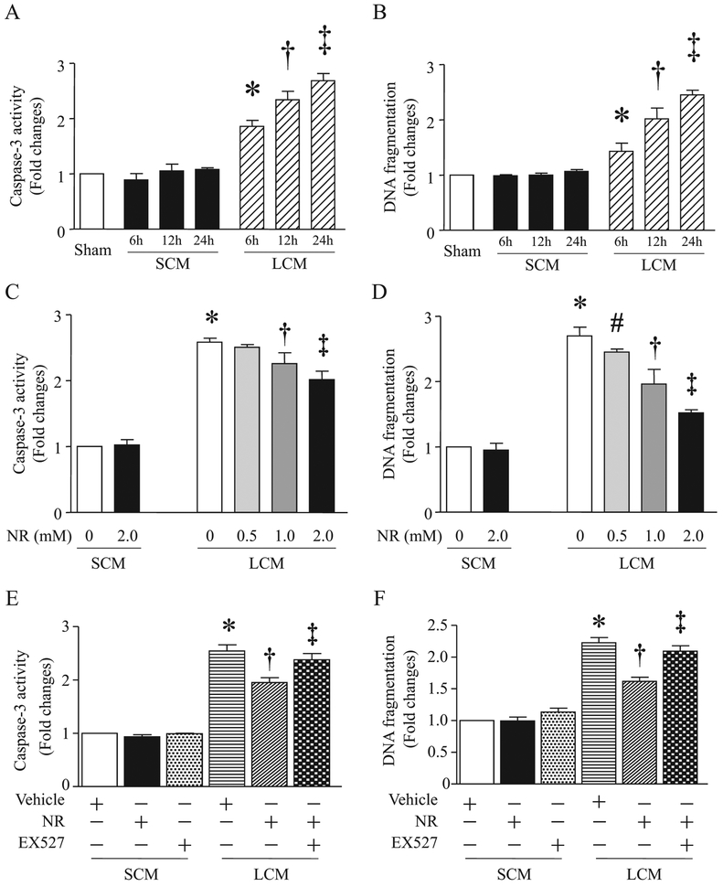
Fig. 7.
Effects of NR on conditioned medium-induced apoptosis in endothelial cells. (A and B) Mouse cardiac microvascular endothelial cells (MCECs) were challenged by a conditioned medium (LCM) from RAW264.7 cells stimulated with LPS or control medium (SCM) from cultured RAW264.7 cells treated with saline for different times. Caspase-3 (A) and DNA fragmentation (B) were analyzed. Data are mean ± SD (n = 3 independent experiments). *P<0.05 vs 0 h, †P<0.05 vs 6 h, ‡P <0.05 vs 12 h. (C and D) MCECs were treated with NR (0.5 mM, 1 mM, 2 mM) or saline for 12 h, and then challenged by LCM or SCM from RAW264.7 cells. Twelve hours after addition of LCM and SCM, caspase-3 activity (C) and DNA fragmentation (D) were measured. Data are mean ± SD (n = 3). *P<0.05 vs 0 + SCM, #P < 0.05 vs 0 + LCM, †P<0.05 vs 0.5 + LCM, ‡P<0.05 vs 1.0 + CM. (E and F) MCECs were pretreated with NR (2 mM) or saline for 12 h, and then stimulated with LCM or SCM in presence of EX527 (1 μM) or vehicle. Twelve hours later, caspase-3 activity (E) and DNA fragmentation (F) were assessed. Data are mean ± SD (n = 3 independent experiments). *P<0.05 vs vehicle + SCM, †P<0.05 vs vehicle + LCM, ‡P<0.05 vs NR + LCM.

